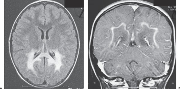
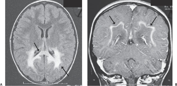
Case 76 A 5-year-old boy with learning delays and gait abnormality. (A) Axial fluid-attenuated inversion recovery (FLAIR) image: there is abnormally high signal in the peritrigonal white matter and corpus callosum (arrows). (B) Coronal T1 post-contrast image: there is linear enhancement along the leading edge of the demyelination adjacent to the subcortical U-fibers (arrows). • X-linked adrenoleukodystrophy: These findings are typical of adrenoleukodystrophy. • Posterior reversible encephalopathy syndrome: This usually affects the cortical and subcortical regions in an asymmetric distribution and has variable patchy enhancement. • Periventricular leukomalacia:
Clinical Presentation
Imaging Findings
Differential Diagnosis
![]()
Stay updated, free articles. Join our Telegram channel

Full access? Get Clinical Tree


