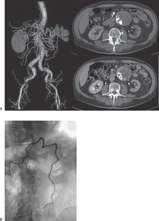
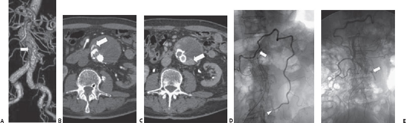
Case 76 An 80-year-old man presents for routine follow-up. (A) Three-dimensional volume-rendered computed tomographic angiogram (CTA) shows stent-graft in place to treat infrarenal aortic aneurysm. The density anterior to the graft (arrow) represents leakage of contrast into excluded aneurysm. (B,C) Arterial and delayed phase axial CT images show contrast within the aneurysm sac (arrows). (D) After selection of the superior mesenteric artery (SMA: arrow), the middle colic artery was subselected, and angiography shows filling of the inferior mesenteric artery (IMA: arrowhead), which supplies the aneurysm. (E) From the SMA approach, the IMA origin is embolized through a microcatheter with coils (arrow).
 Clinical Presentation
 Imaging Findings
 Differential Diagnosis
Stay updated, free articles. Join our Telegram channel

Full access? Get Clinical Tree


