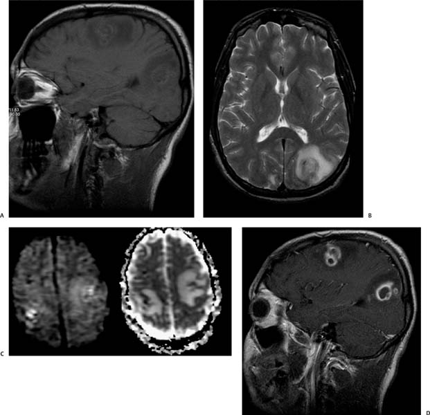
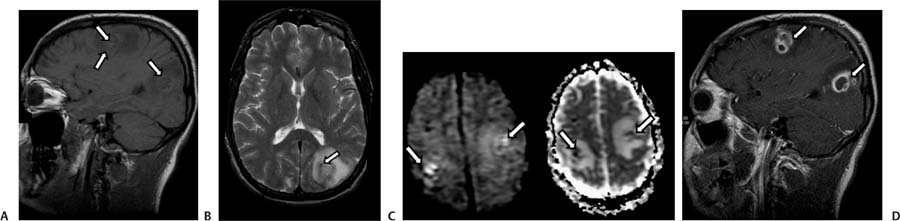
Case 77 A 39-year-old drug addict with heart disease. (A) Sagittal T1-weighted image (WI) shows heterogeneous subcortical nodules (arrows) surrounded by areas of low intensity related to vasogenic edema. The walls demonstrate areas of hyperintensity related to bleeding. (B) Axial T2WI shows vasogenic edema in the left occipital lobe with a central nodular area. There is an area of low signal in the lesion that is related to the bleed (arrow). (C) Diffusion-WI and apparent diffusion coefficient map show bilateral areas of restricted diffusion (arrows). (D) Sagittal T1WI with contrast shows subcortical lesions with ring enhancement (arrows). • Septic emboli:
Clinical Presentation
Imaging Findings
Differential Diagnosis
![]()
Stay updated, free articles. Join our Telegram channel

Full access? Get Clinical Tree


