 Clinical Presentation
A 10-year-old boy with wheezing and dyspnea.
Further Work-up
 Imaging Findings
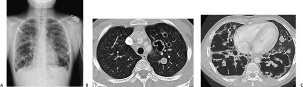
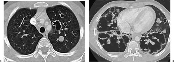
(A) Chest radiograph demonstrates multiple pulmonary nodules and extensive lower lobe–predominant cavities (arrows). (B) Computed tomography (CT) through the upper chest demonstrates a solid 2-cm nodule in the left upper lobe (arrow). Three irregularly shaped cavities are noted anterior to the nodule (arrow). The thickness of the walls is 3 to 4 mm. (C) CT through the lower chest demonstrates posteriorly predominant, large, thick–walled, irregular cavities with extensive parenchymal destruction. An air-fluid level is seen in the largest cavity on the right (arrow). Solid nodules (medial arrow) and a small, thin-walled cyst (lateral arrow) are seen in the anterior left lower lobe and lingula.
Stay updated, free articles. Join our Telegram channel

Full access? Get Clinical Tree


