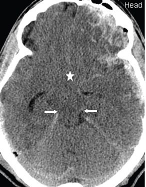


FINDINGS Figures 77-1 and 77-2. Contiguous axial NCCT through the suprasellar cistern. There is effacement of the suprasellar cistern (star) by medially projecting bilateral unci. There is side-to-side compression of the brainstem and obliteration of the perimesencephalic cisterns (arrows). There are bilateral frontal lobe contusions and bilateral fronto-temporal fossa extraaxial hemorrhages.
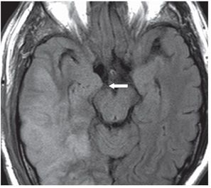
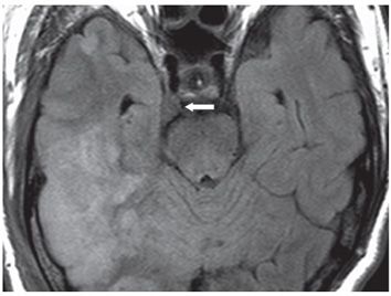
Figures 77-3 and 77-4. Contiguous axial MRI FLAIR through the suprasellar cistern in a different patient with right middle cerebral artery (MCA) territory acute ischemic infarct. There is unilateral right uncal herniation (UH). There is deformity of the right side of the suprasellar star configuration and compression of the right perimesencephalic cistern and midbrain by the shifted right uncus (arrow in Figure 77-3). Figure 77-4 demonstrates the overflow of the right uncus over the free edge of the right tentorium behind the dorsum sella and anterolateral to the upper pons (arrow).
DIFFERENTIAL DIAGNOSIS Bilateral UH, transtentorial herniation (TTH).
Stay updated, free articles. Join our Telegram channel

Full access? Get Clinical Tree


