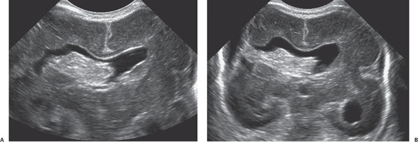
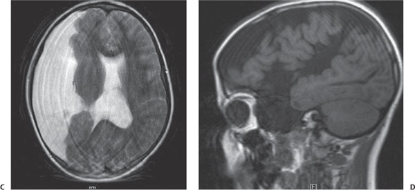
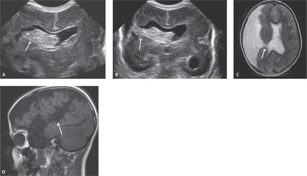
Case 77 A premature newborn with seizures. (A,B) Coronal ultrasound images: There is a cleft extending from the right lateral ventricle through the cortex to the extra-axial fluid. Choroid plexus extends through the cleft (arrows). Note the premature, smooth appearance of the brain and absence of the septum pellucidum. (C) Axial T2 MR image (several years later): again noted is a cleft extending from the lateral ventricle to the extra-axial fluid (arrow). (D) Sagittal T1 MR image (several years later): it is easier to tell on this image that the cleft is lined with gray matter (arrow).

Clinical Presentation
Further Work-up

Imaging Findings

Differential Diagnosis
Stay updated, free articles. Join our Telegram channel

Full access? Get Clinical Tree


