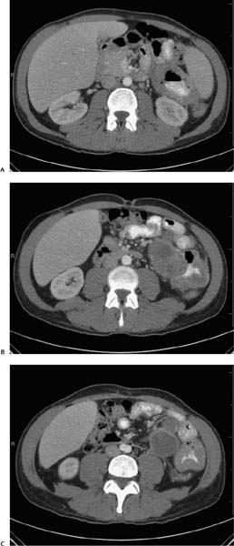
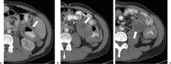
Case 77 A 39-year-old man presents with gastrointestinal bleeding. (A) Contrast-enhanced computed tomography shows marked, circumferential mural thickening (arrow) of the proximal jejunum and no enlarged lymph nodes. (B) More caudal image shows a large exophytic mass with central low-density areas of necrosis or cavitation (arrow). Minimal lymphadenopathy is noted (arrowhead). (C) More caudal image shows two lobulated components of the mass with an enhancing rim and central low density (arrow). • Melanoma: This top diagnostic choice causes cavitary lesions of the small bowel, and metastatic disease is the most common small-bowel neoplasm. • Leiomyosarcoma:
 Clinical Presentation
 Imaging Findings
 Differential Diagnosis
![]()
Stay updated, free articles. Join our Telegram channel

Full access? Get Clinical Tree


