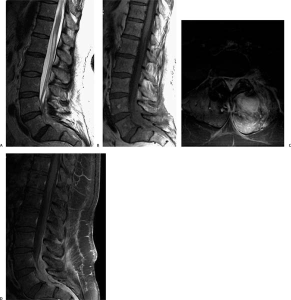
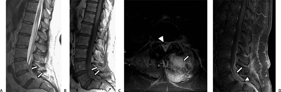
Case 78 A patient with increased radicular pain and fever 1 month after laminectomy. (A) Sagittal T2-weighted image (WI) shows heterogeneous signal in the lower lumbar spine (arrows). (B) Sagittal T1WI shows isointense signal in the lower lumbar spinal canal (arrows); the nerve roots are not seen. x (C) Fat-saturated axial T1WI with contrast shows enhancement of the left paraspinal muscles and the posterior aspect of the spinal canal (arrow) compressing the thecal sac (arrowhead). (D) Fat-saturated T1WI of the lumbar spine with contrast shows concentric anterior and posterior enhancement of the epidural space (arrow) and a central area without enhancement (arrowhead).
Clinical Presentation
Imaging Findings
Differential Diagnosis
Stay updated, free articles. Join our Telegram channel

Full access? Get Clinical Tree


