 Clinical Presentation
A 50-year-old man with sepsis following bone marrow transplant.
Further Work-up

 Imaging Findings

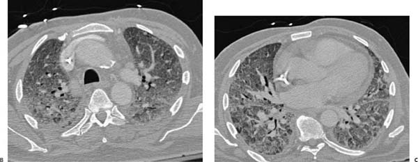
(A) Portable chest radiograph demonstrates an intubated patient with a right internal jugular central venous catheter (arrows). There is diffuse, bilateral pulmonary opacity without evidence of cardiomegaly or pleural effusion. (B) Contrast-enhanced computed tomography (CT; lung window) through the upper chest confirms widespread ground-glass opacity without pleural effusion or frank consolidation (arrows). (C) Contrast-enhanced CT (lung window) through the lower chest shows diffuse ground-glass opacity, mosaic attenuation, and pulmonary edema (arrows). Note the lack of pleural effusions and inhomogeneous distribution.
 Differential Diagnosis
• Acute respiratory distress syndrome (ARDS):
Stay updated, free articles. Join our Telegram channel

Full access? Get Clinical Tree


