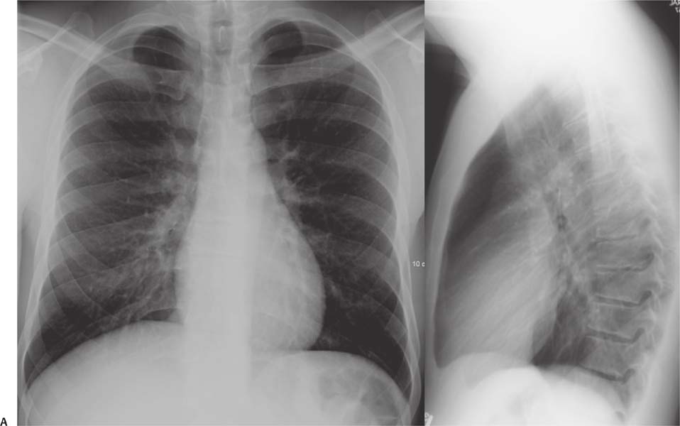
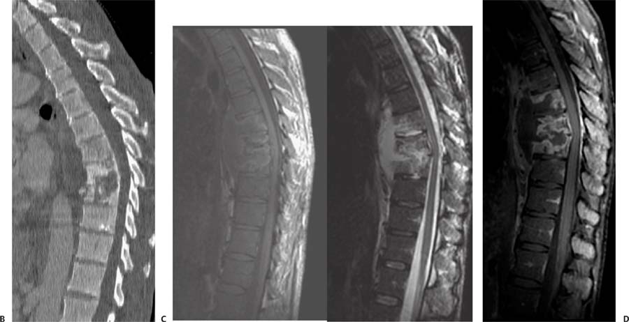
Case 79 A 45-year-old patient with chronic cough who is now experiencing persistent back pain. (A) Lateral radiograph of the thoracic spine shows irregular end plates and narrowed disk spaces of the midthoracic vertebrae (arrows). (B) Sagittal reformatted computed tomography (CT) of the thoracic spine without contrast shows hypodense destructive changes of a vertebra (arrow). There is a prevertebral collection (asterisk). (C) Sagittal T1- and T2-weighted images (WIs) of the thoracic spine show low T1 and high T2 signal of three adjacent vertebral bodies (asterisks). There is an anterior well-defined fluid collection with a thin capsule adjacent to these segments (arrows). (D)
Clinical Presentation
Further Work-up
Imaging Findings
![]()
Stay updated, free articles. Join our Telegram channel

Full access? Get Clinical Tree


