 Clinical Presentation
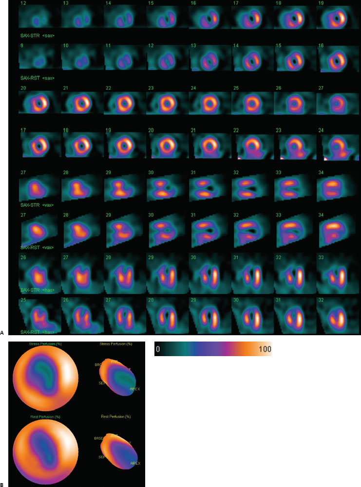
A 68-year-old woman presents with chest pain and undergoes myocardial perfusion scintigraphy at rest and then at stress. The gated images (not shown) demonstrated an apical wall motion abnormality.

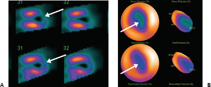
(A) Myocardial perfusion SPECT images demonstrate a medium-sized defect of marked severity in the anterior apex at stress that improves mildly at rest (arrows). Mild aneurysmal dilatation of the apex is also seen. (B) Stress and resting polar maps again demonstrate the severe, slightly reversible defects (arrows).
 Differential Diagnosis
All three diagnoses can demonstrate defects on the rest and stress scans. However, attenuation artifacts should not have regional wall motion abnormalities, and hibernating myocardium requires additional imaging to document.
• Left anterior descending territory myocardial infarction with mild peri-infarct ischemia: This is the most likely diagnosis to account for a resting defect that worsens on stress and has a regional wall motion abnormality.
• Hibernating myocardium:
Related posts:


Stay updated, free articles. Join our Telegram channel

Full access? Get Clinical Tree


