 Clinical Presentation
A 45-year-old woman with severe dyspnea.
Further Work-up
 Imaging Findings
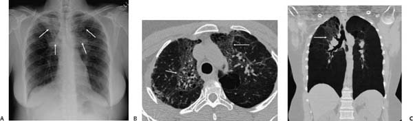
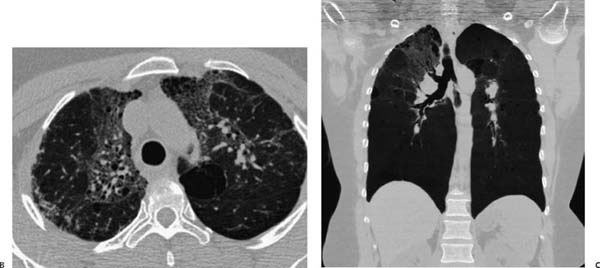
(A) Chest radiograph demonstrates an upper lobe–predominant interstitial abnormality with upper lobe retraction (arrows). There is mild paratracheal and hilar lymphadenopathy. (B) Computed tomography (CT) of the chest (lung windows) demonstrates severe upper lobe volume loss, traction bronchiectasis, and cyst formation (arrows). There is extensive subpleural nodularity. (C) CT of the chest (coronal minimum-intensity projection) demonstrates the striking upper lobe–predominant fibrosis to advantage (arrow).
 Differential Diagnosis
• Sarcoidosis: The striking upper lobe predominance, small subpleural nodules, and lymphadenopathy make end-stage sarcoidosis the best choice.
• Berylliosis: This has a similar imaging appearance, although an appropriate occupational exposure (aerospace, electronics, and ceramics industries) should be elicited. Berylliosis is far less common than sarcoidosis.
Stay updated, free articles. Join our Telegram channel

Full access? Get Clinical Tree


