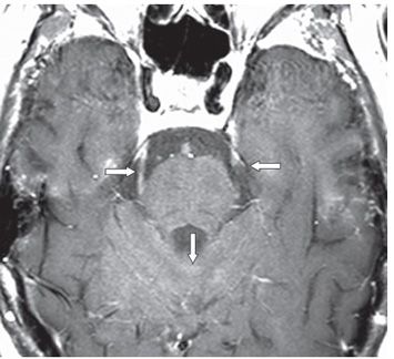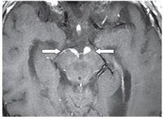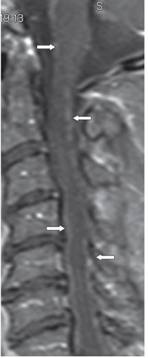


FINDINGS Figure 79-1. Axial post-contrast T1WI through the cerebellopontine angles (CPAs). There is nodular contrast enhancement around the bilateral CN VII and VIII both in the cisternal and in the intracanalicular segments (vertical arrows). There is similar but smaller nodular enhancement along the right CN VI (transverse arrow). Figure 79-2. Axial post-contrast T1WI through the trigeminal nerves. There is nodular but somewhat smooth enhancement along the bilateral trigeminal nerves (transverse arrows). There is also smudgy contrast enhancement around the cerebellar folia (vertical arrow). Figure 79-3. Axial post-contrast T1WI through the interpeduncular cistern. There is nodular and linear contrast enhancement on the surface of the cerebral peduncles (transverse arrows). Figure 79-4. Post-contrast sagittal T1WI through the cervical spine. There is fine nodular enhancement of the surface of the spinal cord and brainstem (arrows). FLAIR images (not shown) demonstrated hyperintensity along CN VII and VIII and surrounding the superior cerebellar folia. Other noncontrast images did not show any significant abnormality other than mild ventriculomegaly.
DIFFERENTIAL DIAGNOSIS
Stay updated, free articles. Join our Telegram channel

Full access? Get Clinical Tree


