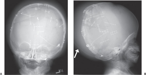
Case 79 A toddler who previously underwent craniotomy and ventriculoperitoneal shunting during resection of a brain tumor presents with headache and a posterior head mass. (A,B) Frontal and lateral radiographs of the skull obtained as part of a ventriculoperitoneal shunt series demonstrate a large, crescentic soft-tissue mass along the back of the skull, as well as disruption of the shunt catheter, a portion of which extends posterior to the mass (arrow). • Disrupted shunt catheter:
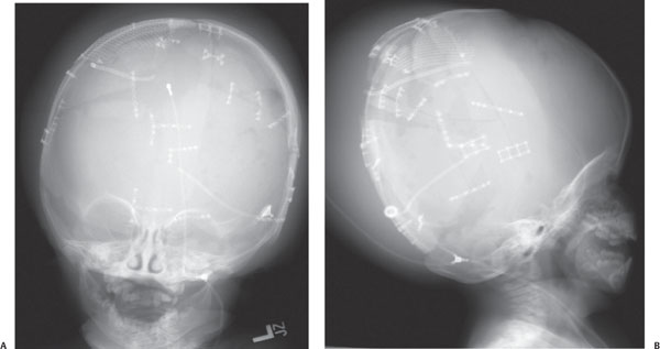
Clinical Presentation
Imaging Findings
Differential Diagnosis
![]()
Stay updated, free articles. Join our Telegram channel

Full access? Get Clinical Tree


