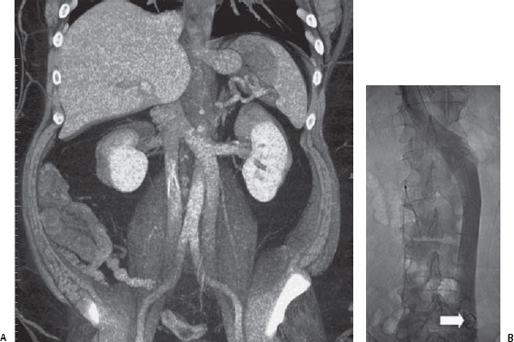
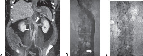
Case 79 A patient presents with gastrointestinal bleeding, deep venous thrombosis (DVT), and recurrent pulmonary embolism (PE) despite caval filtration. (A) Selected image is a coronal volume slab from an infused computed tomographic (CT) scan shows a filter in a right-sided inferior vena cava (IVC: arrow). The IVC is duplicated, with the left component draining into the left renal vein (arrowhead). (B) Conventional venogram with pigtail (arrow) in left component and filter in right component. (C) Fluoroscopic image with bilateral filters in place.
 Clinical Presentation
 Imaging Findings
 Differential Diagnosis
Stay updated, free articles. Join our Telegram channel

Full access? Get Clinical Tree


