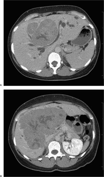
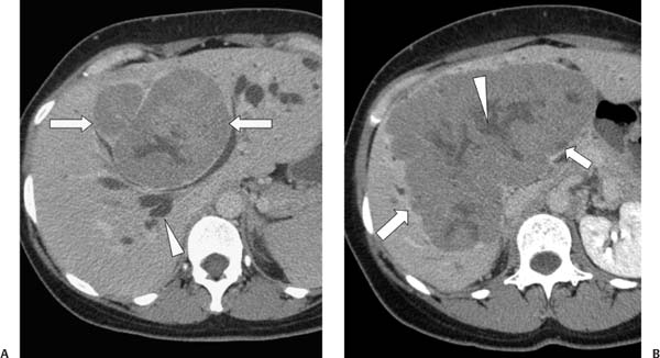
Case 79 A 35-year-old woman presents with right upper quadrant pain. Her α-fetoprotein level is normal. (A) Infused abdominal computed tomography (CT) shows a well-circumscribed, heterogeneous hepatic mass (arrows) that is hypodense to liver parenchyma and obstructs the intrahepatic bile ducts (arrowhead).(B) More caudal image shows the mass to be quite large, with a lobulated contour (large arrow), a dense capsule (small arrow), and a hypodense, radiating central region (arrowhead). • Fibrolamellar carcinoma (FLC): This top choice is suggested by a large, encapsulated mass with a hypodense central scar on delayed imaging and a lobulated contour in a young patient. The α-fetoprotein level is typically normal, as in this case. • Hepatic adenoma:
 Clinical Presentation
 Imaging Findings
 Differential Diagnosis
![]()
Stay updated, free articles. Join our Telegram channel

Full access? Get Clinical Tree


