CASE 79
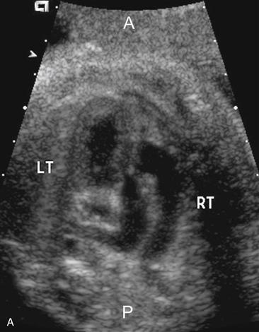
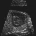
A = anterior; P = posterior; LT = left; RT = right.
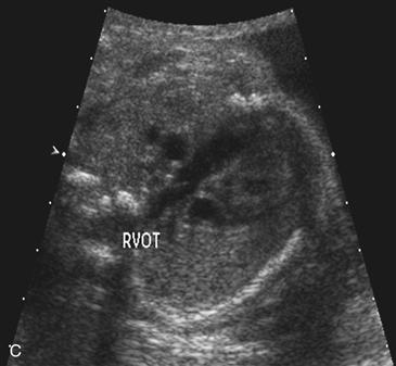
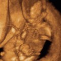
Used with permission from McGahan JP, et al. Fetal Heart. In McGahan JP, Goldberg B [eds]: Diagnostic Ultrasound, 2nd ed. New York, NY. Informa Healthcare USA, 2008; 1269.
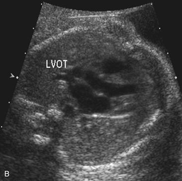
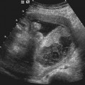
Used with permission from McGahan JP, et al. Fetal Heart. In McGahan JP, Goldberg B [eds]: Diagnostic Ultrasound, 2nd ed. New York, NY. Informa Healthcare USA, 2008; 1269.
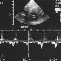
History: A patient with a family history of congenital heart disease presents at 24 weeks’ gestation for evaluation.
1. What should be included in the differential diagnosis for the imaging finding presented in Figure A? (Choose all that apply.)
C. Double outlet of the right ventricle
D. Dextraposed transposition of the great arteries (d-TGA)
2. What is the most common associated defect with d-TGA?
B. VSD
D. Tricuspid or mitral valve atresia
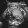
3. Which one of the following is not an important finding used to help identify potential cases of d-TGA?
A. Abnormal cardiac axis of approximately 60 degrees on four-chamber view of the heart
B. Presence of VSD on four-chamber view of the heart
Stay updated, free articles. Join our Telegram channel

Full access? Get Clinical Tree


