Case 79
Case History
A 49-year-old woman presents with new left subareolar calcifications on her screening mammogram.
Physical Examination
• normal exam
Mammogram
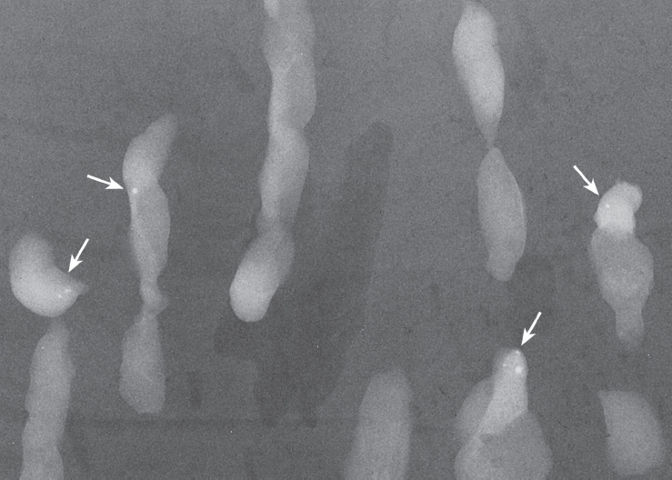
Calcifications (Figs. 79–1 and 79–2)
• type: punctate
• distribution: regional
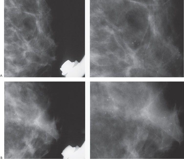
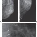
Figure 79–1. Numerous round punctate calcifications are present in the subareolar region. (A). Left MLO magnification mammogram. (B). Left CC magnification mammogram.
