Case 8 A 2-year-old girl presenting with developmental delay and seizures. (A) Axial T1-weighted image (WI) shows severely decreased sulcation of the brain with a thick cortex (arrows). (B) Sagittal T1WI shows a thick cortical layer with severely decreased sulcation. (C) Axial T2WI shows severely decreased sulcation of the brain with a thick cortex (arrows). (D) Axial T2WI shows severely decreased sulcation of the brain with a thick cortex (arrows). • Type I (classic) lissencephaly:
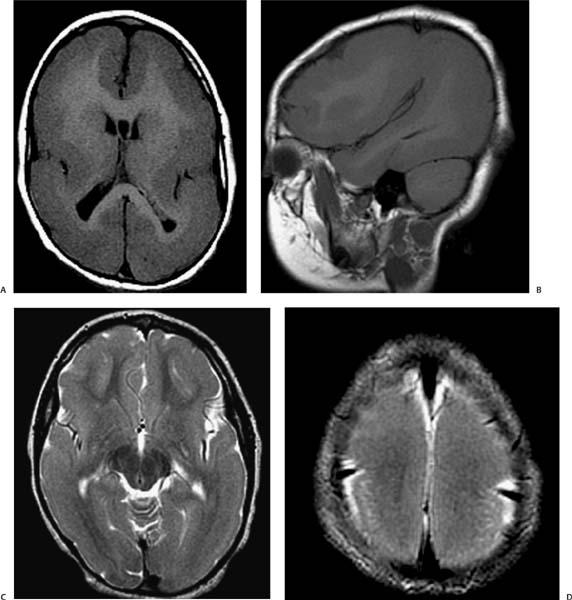
Clinical Presentation
Imaging Findings
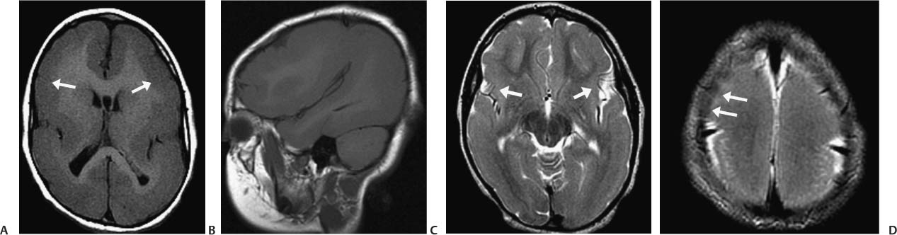
Differential Diagnosis
![]()
Stay updated, free articles. Join our Telegram channel

Full access? Get Clinical Tree


