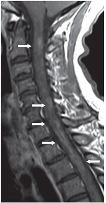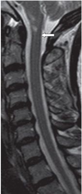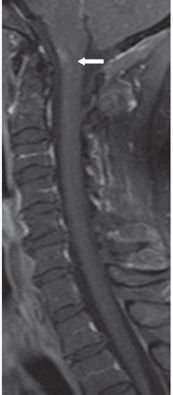


FINDINGS Figure 8-1. Sagittal short tau inversion recovery (STIR) cervical spine MRI demonstrating extensive continuous spinal cord hyperintensity from C1 to T5. The thoracic spine study (not shown) showed inferior extension of the lesion into the thoracic spinal cord. Figure 8-2. Sagittal post-contrast T1WI at the same time showing extensive patchy contrast enhancement throughout the lesion (arrows). Figures 8-3 and 8-4. Sagittal T2WI and post-contrast sagittal T1WI, respectively, 8 weeks later showing healing of the extensive lesion with only a small residual spinal cord lesion with contrast enhancement anteriorly at the foramen magnum level (arrows).
DIFFERENTIAL DIAGNOSIS Transverse myelitis, hypertensive myelopathy, ependymoma, astrocytoma.
DIAGNOSIS Longitudinally extensive transverse myelitis (LETM).
DISCUSSION
Stay updated, free articles. Join our Telegram channel

Full access? Get Clinical Tree


