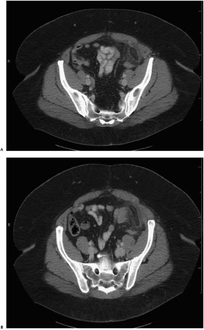
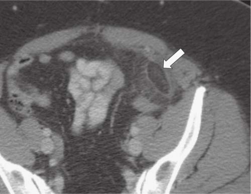
Case 8 A 50-year-old woman presents to the emergency department with the recent onset of left lower quadrant pain. Enhanced axial computed tomography (CT) shows a fatty structure anterior to the sigmoid colon surrounded by a hyperdense rim (arrow) and edematous fat stranding. A central focus of increased density within this structure represents infarcted vein due to torsion of the vascular pedicle of the epiploic appendage, which is the root cause of this condition. • Acute epiploic appendagitis: This usually presents as an oval fatty structure adjacent to the anterior sigmoid colon surrounded by a hyperdense ring. • Omental infarction: This can also present as a larger structure in the right lower quadrant with a heterogeneous whirl of stranded fat between the abdominal wall and ascending/transverse colon. However, it displays no hyperdense ring in most cases.
 Clinical Presentation
 Imaging Findings
 Differential Diagnosis
Stay updated, free articles. Join our Telegram channel

Full access? Get Clinical Tree


