Case 8

 Clinical Presentation
A 73-year-old man with suprapubic pain and hematuria.
 Imaging Findings

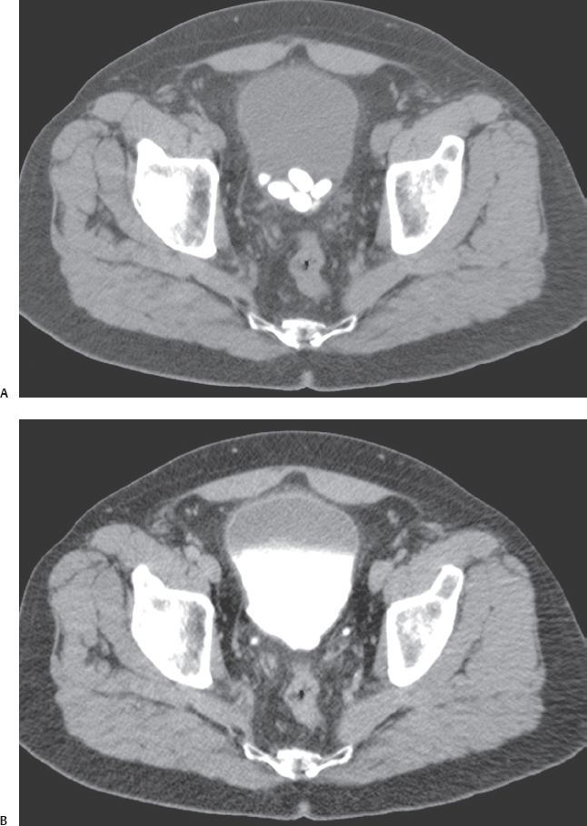
(A) Precontrast axial computed tomography (CT) image through the pelvis shows multiple calcified oval and round structures within the dependent portion of the lumen of the urinary bladder (arrow). (B) Postcontrast axial CT image obtained in the excretory phase at the same level as Figure A shows that the urinary bladder lumen is filled with excreted contrast (asterisk). The calcifications seen in the precontrast phase have been obscured by the luminal contrast. The bladder wall is normal. No filling defect is seen. No diverticula are present.
 Differential Diagnosis
• Urinary bladder stones: Round or oval, smooth calcified structures in the lumen of the urinary bladder are characteristic of urinary bladder stones. They lie in the dependent portion of the bladder.
• Foreign bodies in urinary bladder: These are mostly fragments of various urologic catheters and tubes or sutures. They are seldom round and smooth to start with. However, they may act as nidi for subsequent stone formation.
• Urinary bladder neoplasm:
Stay updated, free articles. Join our Telegram channel

Full access? Get Clinical Tree


