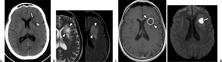
Case 80 A 36-year-old woman with a history of heroin use presenting with diffuse headache, fever, and seizures. (A) Axial computed tomography (CT) without contrast shows a ring isodense to the gray matter (arrowhead) in the left frontal lobe. The lesion, located at the gray–white matter junction, has a hypodense center and is surrounded by vasogenic edema (arrow). (B) Axial T2-weighted image (WI) and fluid-attenuated inversion recovery image show the left frontal lesion with a rim of low intensity (arrowheads) and peripheral vasogenic edema. (C) Axial T1WI with contrast shows a round, well-defined, ring-enhancing lesion in the left frontal lobe (arrowheads). The inner and outer margins are smooth. (D) Diffusion-WI shows a “lightbulb” sign of the left frontal lesion (arrow). • Cerebral abscess:
Clinical Presentation
Further Work-up
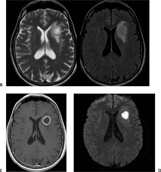
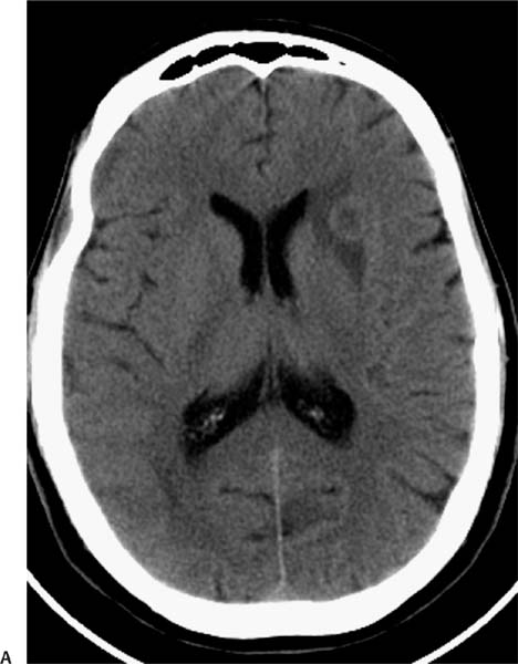
Imaging Findings
Differential Diagnosis
![]()
Stay updated, free articles. Join our Telegram channel

Full access? Get Clinical Tree


