 Clinical Presentation
A 40-year-old woman with fever and an elevated white cell count.
Further Work-up

 Imaging Findings
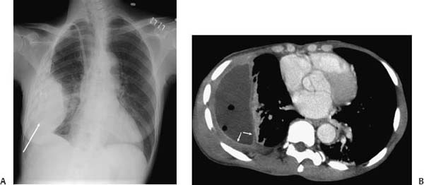
(A) Posteroanterior chest radiograph demonstrates a large pleura-based mass on the right (arrow). A hemodialysis catheter is in the expected location. (B) Contrast-enhanced computed tomography (CT; soft-tissue windows) demonstrates a loculate pleural effusion containing air. The pleura is thickened and hyperdense (arrows).
 Differential Diagnosis
• Empyema: Pleural thickening and enhancement surrounding a pleural effusion are highly suggestive of an empyema.
• Mesothelioma:
Stay updated, free articles. Join our Telegram channel

Full access? Get Clinical Tree


