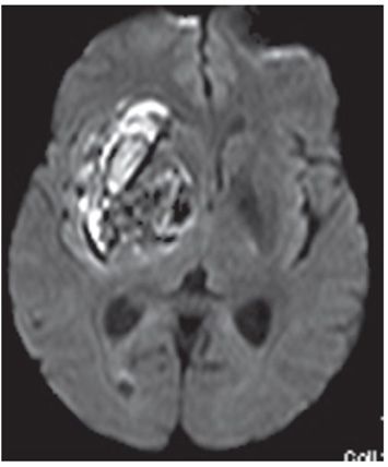
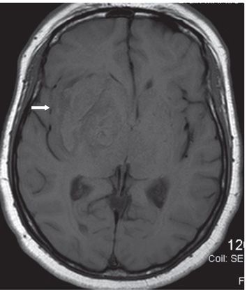
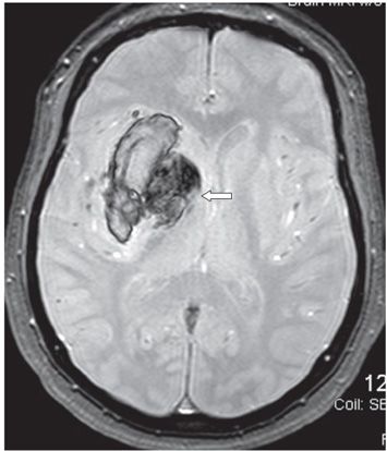
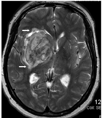
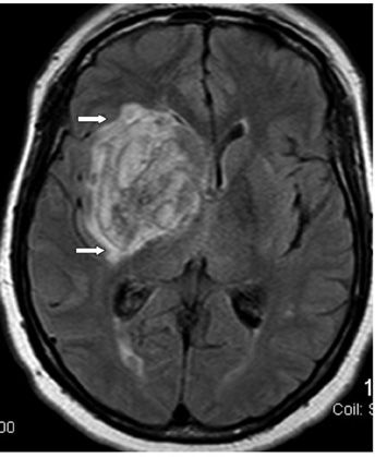
FINDINGS Figure 80-1. Axial NCCT through the basal ganglia. There are two hyperdense masses in the right basal ganglia, one in the right globus pallidus (transverse arrow) and the larger lateral one straddles the putamen and external capsule (vertical arrow). Both communicate superiorly (not shown). There is effacement of the right frontal horn due to mass effect. Figure 80-2. Axial DWI through the basal ganglia. There is heterogeneous area of restricted diffusion in the right basal ganglia. Figure 80-3. Axial T1WI through the basal ganglia. The right basal ganglia mass is mainly isointense with thin surrounding hypointense halo. There is effacement of the right sylvian fissure (arrow). Figure 80-4. Axial GRE through the basal ganglia. There is heterogeneous blooming of the mass (arrow). Figures 80-5 and 80-6. Axial T2WI and FLAIR through the mass. The mass has similar heterogeneous pattern of mixed hyperintensity and isointensity with local mass effect and midline shift. There is mild surrounding hyperintensity consistent with vasogenic edema (arrows).
DIFFERENTIAL DIAGNOSIS N/A.
DIAGNOSIS
Stay updated, free articles. Join our Telegram channel

Full access? Get Clinical Tree


