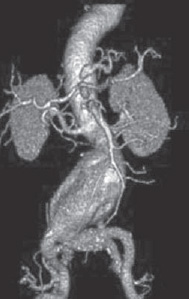
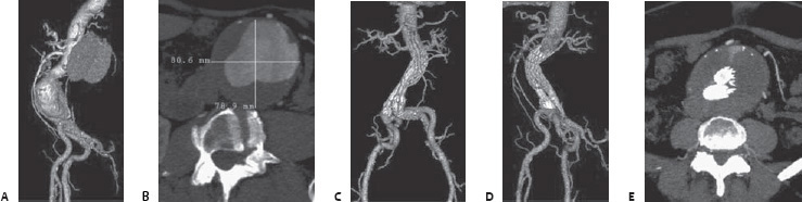
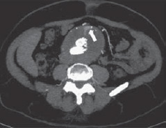
Case 80 The patient is a 50-year-old who underwent repair of an abdominal aortic aneurysm (AAA). Case images: Frontal and lateral three-dimensional (3D) volume-rendered computed tomographic angiograms (CTAs) show a large infrarenal AAA extending to the iliac bifurcation. The axial source image most accurately reflects the true diameter of 8 cm. Frontal and lateral 3D volume-rendered CTAs show interval placement of an aortic endograft. Axial image shows contrast leakage (arrow) into the anterior aneurysm sac at the level of the inferior mesenteric artery.
 Clinical Presentation
 Imaging Findings
 Differential Diagnosis
 Essential Facts
Stay updated, free articles. Join our Telegram channel

Full access? Get Clinical Tree


