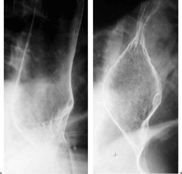
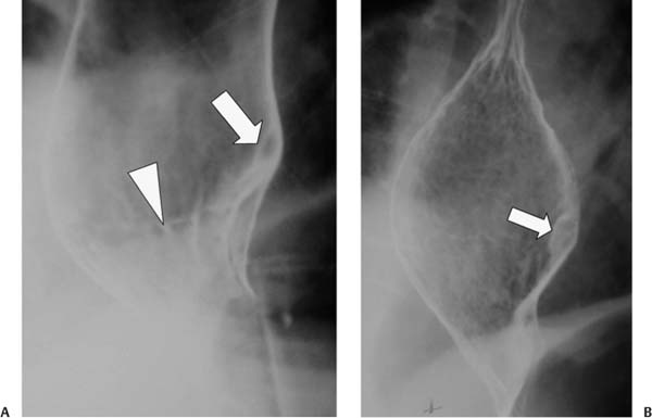
Case 80 A 35-year-old man presents to the gastroenterology clinic with chronic, mild midepigastric pain. (A) Double-contrast esophagogram shows a prominent squamocolumnar mucosal junction (Z line; arrowhead) and a small submucosal mass (arrow) at this level. (B) The mass (arrow) is well circumscribed and makes obtuse angles with the esophageal wall, identifying it as submucosal. • Leiomyoma: This is the most likely diagnosis for a smoothly marginated, submucosal (intramural) esophageal mass, based purely on statistical probability. The imaging findings are nonspecific. • Neural tumor: Schwannoma, neurofibroma, or granular cell tumor may be submucosal. Neurofibromas typically occur in patients with von Recklinghausen disease.
 Clinical Presentation
 Imaging Findings
 Differential Diagnosis
Stay updated, free articles. Join our Telegram channel

Full access? Get Clinical Tree


