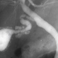
CASE 80
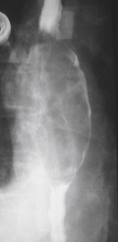
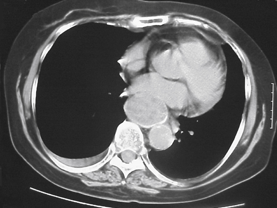
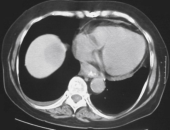
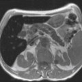
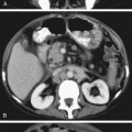
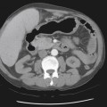
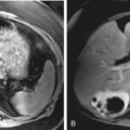
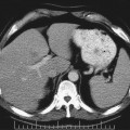
History: A 64-year-old woman presents with dysphagia and hematemesis.
1. Which of the following should be included in the differential diagnosis of the imaging finding shown in the figures? (Choose all that apply.)
2. What tumor most often invades the esophagus through direct extension?
3. What is the most common cause of hematogenous metastases to the esophagus?
Stay updated, free articles. Join our Telegram channel

Full access? Get Clinical Tree


