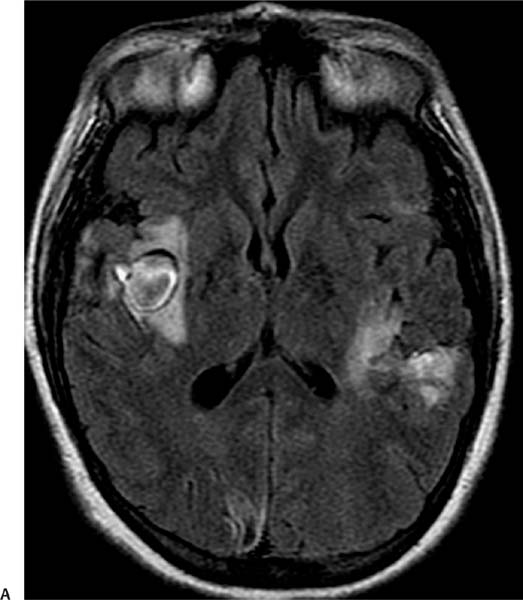
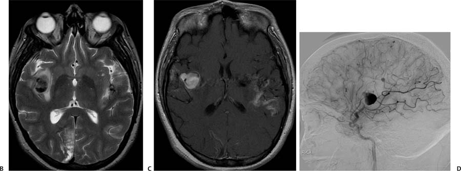
Case 81 A 35-year-old drug addict with bacterial endocarditis presenting with the sudden onset of severe, worsening headache. (A) Axial fluid-attenuated inversion recovery image shows a round lesion in the deep sylvian fissure on the right (black arrow) surrounded by vasogenic edema. The lesion has a well-defined capsule of low intensity. Additional hyperintensities are seen in the perisylvian cortex on the left and in the right occipital lobe (white arrows). (B) Axial T2-weighted image (WI) shows a hypointense round lesion in the deep sylvian fissure on the right (white arrow) and a smaller contralateral lesion (black arrow). (C) Axial T1WI with contrast shows diffuse enhancement of the right sylvian lesion (white arrow). There is cortical enhancement in the cortex surrounding the left sylvian fissure (black arrow) and in the left occipital lobe. (D)
Clinical Presentation
Further Work-up
Imaging Findings
![]()
Stay updated, free articles. Join our Telegram channel

Full access? Get Clinical Tree


