 Clinical Presentation
A 32-year-old woman with progressive dyspnea.
Further Work-up
 Imaging Findings
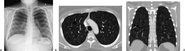
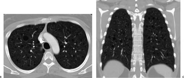
(A) Chest radiograph is normal. (B) Computed tomography (CT) of the chest (axial, lung windows) shows diffusely scattered thin-walled cysts (arrows). The intervening lung is normal. (C) CT of the chest (coronal, lung windows) demonstrates that the cysts are diffusely scattered and affect the lung bases (arrows).
 Differential Diagnosis
Stay updated, free articles. Join our Telegram channel

Full access? Get Clinical Tree


