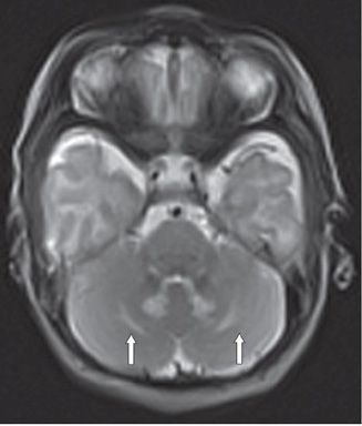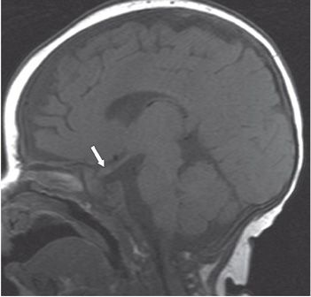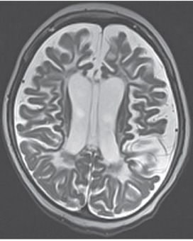


FINDINGS Figure 81-1. Axial T2WI through the corona radiata shows symmetrical bilateral deep hemispheric white matter (WM) hyperintensity (stars). Figure 81-2. Axial T2WI through the cerebellum in a companion case. There is bilateral medial cerebellar (dentate nuclei) symmetrical hyperintensity (arrows). Figure 81-3. Parasagittal T1WI in another companion case shows enlarged optic nerve (arrow). Figure 81-4. Axial T2WI in an end-stage patient. There is marked cerebral volume loss and hyperintense atrophic WM.
DIFFERENTIAL DIAGNOSIS
Stay updated, free articles. Join our Telegram channel

Full access? Get Clinical Tree


