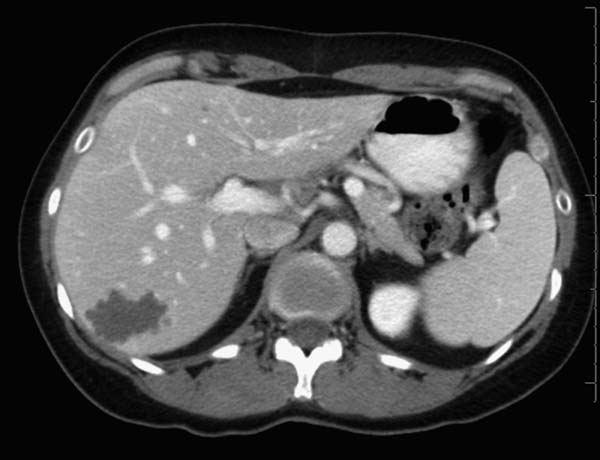
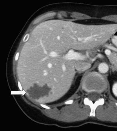
Case 81 A 45-year-old woman presents with right upper quadrant pain without fever or leukocytosis. Contrast-enhanced computed tomography (CT) shows a well-circumscribed, multiloculated cystic lesion (arrow) in the right lobe of the liver. No enhancing nodular components are visible. Scattered, smaller low-density foci are seen in the liver, most likely cysts. • Biliary cystadenoma: This is the strongest contender as the diagnosis for a multiloculated, smoothly marginated cystic lesion in the liver without clinical signs of infection. • Hepatic cyst: This can have a similar appearance in a minority of cases. This patient has other low-density lesions suspicious for simple cysts. • Echinococcal cyst: This often results from infection by Echinococcus granulosus or Echinococcus multilocularis
 Clinical Presentation
 Imaging Findings
 Differential Diagnosis
![]()
Stay updated, free articles. Join our Telegram channel

Full access? Get Clinical Tree


