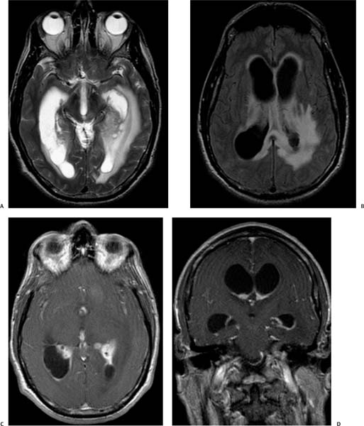
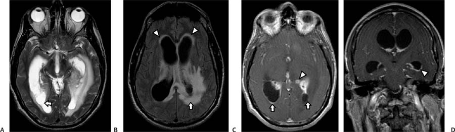
Case 82 A 38-year-old man with severe headache and fever. (A) Axial T2-weighted image (WI) shows dilated lateral ventricles with a septum in the right occipital horn (arrow). (B) Axial fluid-attenuated inversion recovery (FLAIR) image shows hyperintense signal around the ventricles (arrowheads) that is more prominent around the right atrium (arrow). (C) Axial T1WI of the brain with contrast shows enhancement at the ependymal lining (arrows). There is an enhancing nodule (arrowhead) in the left thalamic region. (D) Coronal T1WI with contrast shows enhancement of the ventricular wall (arrowhead). Note the dilatation of the lateral ventricles. • Ventriculitis:
Clinical Presentation
Imaging Findings
Differential Diagnosis
![]()
Stay updated, free articles. Join our Telegram channel

Full access? Get Clinical Tree


