 Clinical Presentation
A 32-year-old man who is a smoker with fatigue and chronic cough.
 Imaging Findings

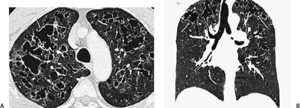
(A) Computed tomography (CT) of the chest (axial, lung windows) shows bilateral irregularly shaped cysts and scattered nodules (arrows). (B) CT of the chest (coronal re-formation) demonstrates upper lobe predominance. The lung bases are not affected (arrows).
 Differential Diagnosis
• Pulmonary Langerhans cell histiocytosis (PLCH): Irregularly shaped, upper lobe–predominant cysts with variable wall thickness and scattered indistinct nodules in a young male smoker make PLCH the best choice.
• Lymphangioleiomyomatosis (LAM):
Stay updated, free articles. Join our Telegram channel

Full access? Get Clinical Tree


