With permission from Applied Neurology UBM Medica. Case previously published in Appl Neurol. 2007;3:35–37.


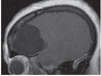
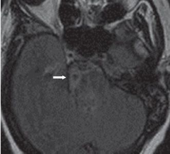
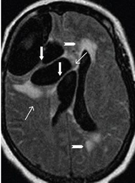
FINDINGS Figures 82-1 and 82-2. Axial and sagittal post-contrast T1WI, respectively, through the frontal lobe showing a huge multiloculated cerebrospinal fluid (CSF) intensity cyst in the right frontal lobe with compression of the right lateral ventricle. The cyst is surrounded by a very thin non-contrast-enhancing wall and apparently communicates with the right frontal horn (arrow). There is midline shift from right to left as demonstrated in Figure 82-1. Figure 82-3. Axial FLAIR image through the pons showing a right perimesencephalic or pontine cyst (possibly on the surface of the pons) with surrounding somewhat thick T2 hyperintensity (arrow) presumably arachnoid inflammation and/or pontine gliosis or edema. Figure 82-4. Axial FLAIR through the giant cyst in the right frontal lobe showing internal septations (arrows), midline shift from right to left, surrounding confluent T2 hyperintensity anteromedially and posteriorly (line arrows) consistent with edema or gliosis. There is also confluent T2 hyperintensity around the dilated left lateral ventricle frontal and occipital horns (chevrons) consistent with transependymal CSF permeation. Lineal hyperintensities are present in the subarachnoid spaces consistent with meningitis.
DIFFERENTIAL DIAGNOSIS
Stay updated, free articles. Join our Telegram channel

Full access? Get Clinical Tree


