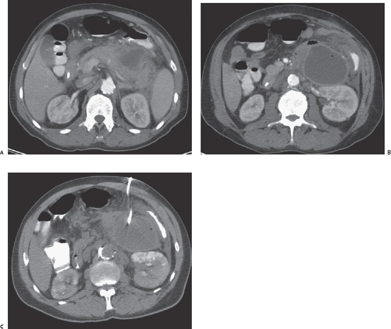
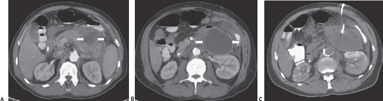
Case 82 A 46-year-old man presents to the emergency department with fever, nausea, vomiting, and back pain. The image in Figure A was obtained on the day of admission. The images in Figures B and C were obtained 4 weeks later. (A) Infused computed tomographic (CT) scan shows fluid collection (arrows), fat stranding, and an enlarged pancreas, consistent with acute fluid collection of pancreatitis. (B) Image obtained 4 weeks later shows enlarging, painful pseudocyst with a well-defined, enhancing wall (arrows). (C) A percutaneous drain was placed.
 Clinical Presentation
 Imaging Findings
 Differential Diagnosis
Stay updated, free articles. Join our Telegram channel

Full access? Get Clinical Tree


