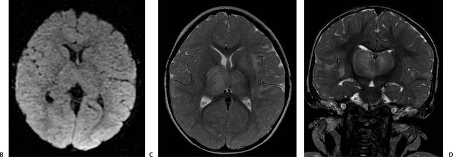
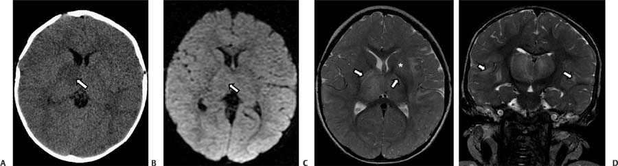
Case 83 A 1-year-old with altered mental status and seizures following a viral illness. (A) Axial computed tomography (CT) shows low attenuation in the right thalamus and posterior limb of the internal capsule (arrow). (B) Axial Diffusion-weighted image (WI) fails to show restriction in the right thalamus (arrow). (C) Axial T2WI demonstrates increased signal intensity in the thalami, internal capsules (arrows), lenticular nucleus on the left (asterisk), and subcortical white matter bilaterally. (D) Coronal T2WI shows enlargement and increased signal in the thalami. There is also increased signal in the subcortical white matter bilaterally (arrows). • Acute disseminated encephalomyelitis (ADEM):
Clinical Presentation
Further Work-up
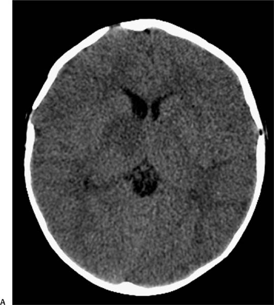
Imaging Findings
Differential Diagnosis
![]()
Stay updated, free articles. Join our Telegram channel

Full access? Get Clinical Tree


