 Clinical Presentation
A 15-year-old boy with worsening dyspnea.
Further Work-up
 Imaging Findings
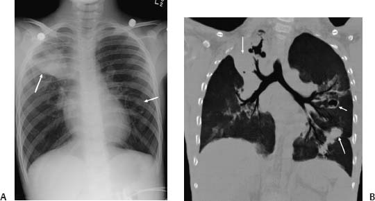
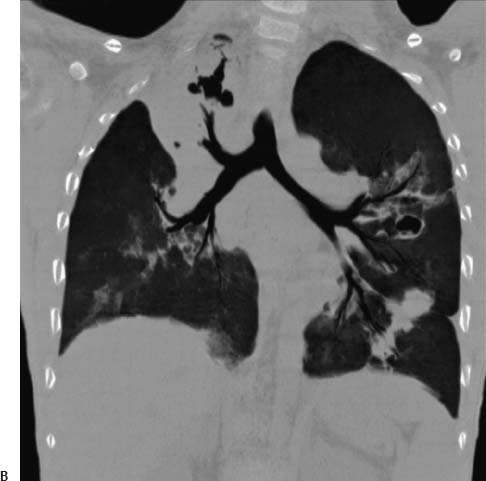
(A) Chest radiograph demonstrates multiple pulmonary nodules and a cavitary nodule in the left lung (arrows). (B) Computed tomography of the chest (coronal re-formation) shows the right upper lobe consolidation to advantage. The cavitary nodule in the left upper lobe has a thin but irregular wall. Solid nodules are also seen in the left lower lobe (arrows).
 Differential Diagnosis
Stay updated, free articles. Join our Telegram channel

Full access? Get Clinical Tree


