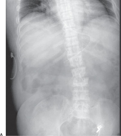
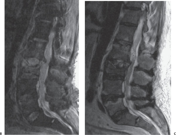
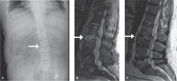
Case 83 A child with painful scoliosis. (A) Frontal abdominal radiograph demonstrates levoscoliosis of the lumbar spine associated with narrowing of the L2-3 intervertebral disk space and possible destruction of the end plates of the adjacent vertebrae (arrow). (B,C) Sagittal T2 and T1 post-contrast magnetic resonance images (MRIs) of the lumbar spine demonstrate destruction of the intervertebral disk and adjacent vertebral end plates with abnormally increased signal and enhancement (arrows), as well as herniation of the disk contents into the vertebral canal. • Diskitis: The plain radiographic findings of intervertebral disk space narrowing and end plate irregularity, as well as MRI findings of disk and end plate inflammation and destruction, are diagnostic. • Neoplasm:
Clinical Presentation
Further Work-up
Imaging Findings
Differential Diagnosis
![]()
Stay updated, free articles. Join our Telegram channel

Full access? Get Clinical Tree


