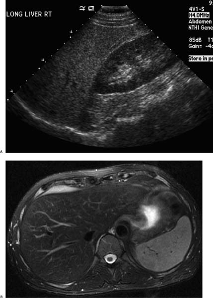
Case 83 A 54-year-old man presents for further work-up of an incidental finding on abdominal ultrasound. (A) Ultrasound shows diffuse, coarse echogenicity of the liver parenchyma (arrowhead) compared with the normal-appearing kidney parenchyma (arrow).(B) T2-weighted magnetic resonance imaging (MRI) shows a homogeneously hypointense liver (arrow) compared with the normal-appearing spleen (arrowhead). • Hemochromatosis (primary): The top diagnosis, it classically causes markedly decreased signal intensity (SI) on T2-weighted MRI and a coarsely echogenic liver on ultrasound due to hepatic fibrosis. • Fatty infiltration of the liver:
 Clinical Presentation
 Imaging Findings
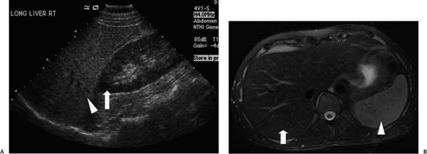
 Differential Diagnosis
![]()
Stay updated, free articles. Join our Telegram channel

Full access? Get Clinical Tree


