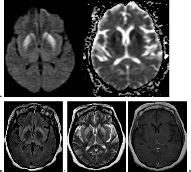
Case 84 A 45-year-old woman with rapidly progressive dementia. (A) Axial Diffusion-weighted image (WI) and apparent diffusion coefficient map show areas of restriction in the caudate head and lenticular nuclei (arrowheads). (B) Axial fluid-attenuated inversion recovery magnetic resonance imaging (MRI) shows symmetric areas of increased T2 signal in the basal ganglia and thalami (asterisks). (C) Axial T2WI shows symmetric areas of increased T2 signal in the basal ganglia as well as in the thalami (asterisks). (D) Axial T1WI after gadolinium injection shows no enhancement (arrow). • Creutzfeldt-Jakob disease (CJD): Symmetric areas of restricted diffusion and T2 hyperintensity involving the basal ganglia in a patient without acute alteration of mental status are highly suggestive of CJD. The lesions do not show mass effect or enhancement. • Hypoxic ischemic injury:
Clinical Presentation
Imaging Findings
Differential Diagnosis
![]()
Stay updated, free articles. Join our Telegram channel

Full access? Get Clinical Tree


