 Clinical Presentation
A 55-year-old woman with Sjögren disease and chronic dyspnea.
 Imaging Findings
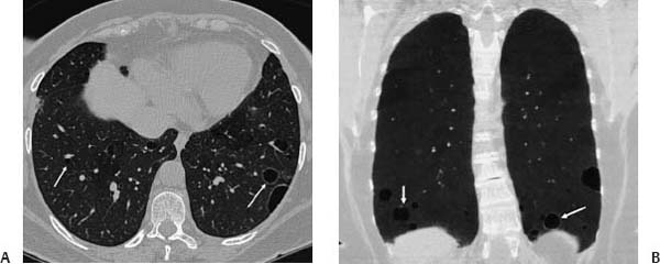
(A) Computed tomography (CT) of the chest (lung windows) at the lung bases shows randomly distributed thin-walled cysts (arrows). There are scattered small centrilobular nodules, minimal ground-glass opacity, and mild bronchial wall thickening. (B) CT of the chest (coronal minimum-intensity projection) shows the distribution of the thin-walled cysts to advantage (arrows).
 Differential Diagnosis
• Lymphoid interstitial pneumonia (LIP):
Stay updated, free articles. Join our Telegram channel

Full access? Get Clinical Tree


