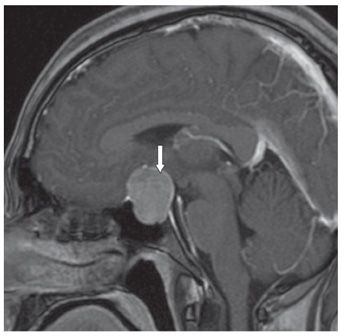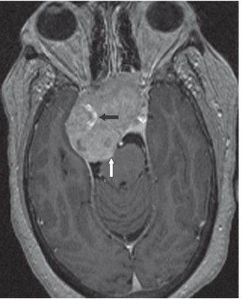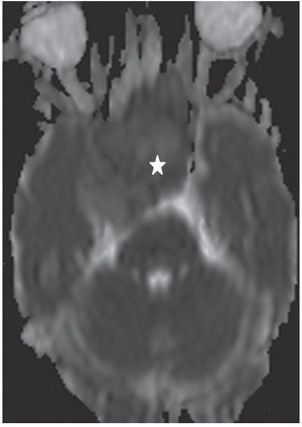


FINDINGS Figure 84-1. Coronal post-contrast T1WI through the sella turcica. There is a 2.5-cm enhancing sella/suprasellar mass (arrows) without cavernous sinus extension. The mass compresses the optic chiasm that cannot be identified. Figure 84-2. Sagittal post-contrast T1WI. There is expanded sella turcica. The mass is compressing the anterior aspect of the third ventricle (arrow). There is no hydrocephalus. Figure 84-3. Axial post-contrast T1WI through the sella in a companion case. There is a large sella-enhancing mass extending into the right cavernous sinus, encasing the internal carotid artery (arrow) and compressing the right temporal lobe. There is posterior extension compressing the pons (vertical arrow). Figure 84-4
Stay updated, free articles. Join our Telegram channel

Full access? Get Clinical Tree


