Case 84
Case History
A 72-year-old woman developed a cluster of calcifications that increased in number when followed at 6-month intervals for 1 year.
Physical Examination
• normal exam
Mammogram
Calcifications (Figs. 84–1 and 84–2)
• type: punctate
• distribution: grouped/clustered
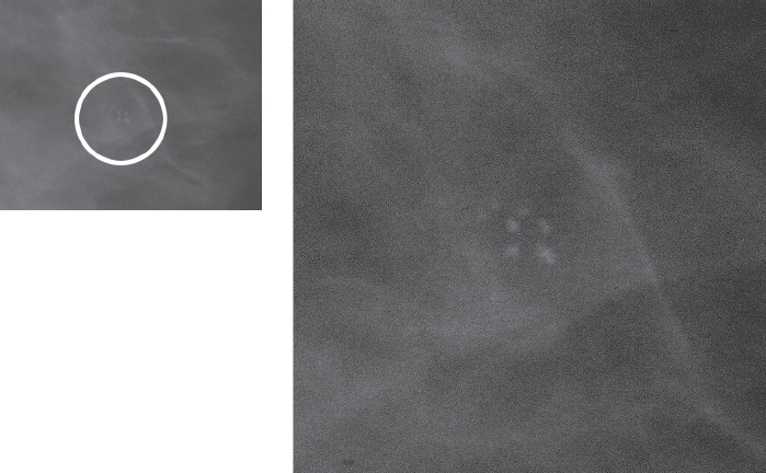
Figure 84–1. Left MLO magnification mammogram: A cluster of fairly uniform punctate calcifications is present inferior to the nipple (circle). No associated mass is appreciated on the mammogram.
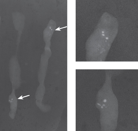
Figure 84–2. Specimen radiograph of 11 gauge core biopsy samples of the calcifications identified in Figure 84–1: In addition to the smooth, fairly round calcifications, there are numerous, smaller, fainter, punctate micro calcifications (arrows). These small calcifications cannot be appreciated on the prebiopsy magnification view (Fig. 84–1).
Ultrasound
Low Frequency


Stay updated, free articles. Join our Telegram channel

Full access? Get Clinical Tree


