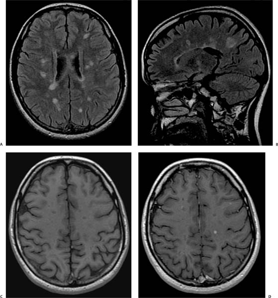
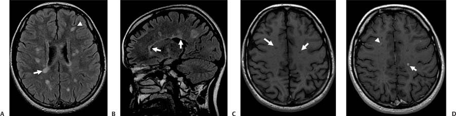
Case 85 A 30-year-old with headache, weakness, and optic neuritis. (A) Axial fluid-attenuated inversion recovery (FLAIR) image shows numerous round and oval areas of increased signal in the periventricular (arrow) and subcortical (arrowhead) white matter. (B) Sagittal FLAIR image demonstrates involvement of the transcallosal white matter tracts (arrows). (C) Axial T1-weighted image (WI) shows low T1 signal (arrows) in some of the lesions. (D) Axial T1WI after gadolinium administration shows peripheral (arrowhead) and central (arrow) enhancement in some of the lesions. • Multiple sclerosis (MS): Characteristic MS lesions are periventricular in location and ovoid in shape, and they may or may not show enhancement, which is often ringlike. Lesions in the corpus callosum, the brainstem beyond the pons, and the spinal cord increase the specificity for MS. • Chronic small-vessel ischemic disease:
Clinical Presentation
Imaging Findings
Differential Diagnosis
![]()
Stay updated, free articles. Join our Telegram channel

Full access? Get Clinical Tree


