 Clinical Presentation
A 67-year-old woman with chronic cough.
Further Work-up
 Imaging Findings
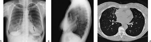
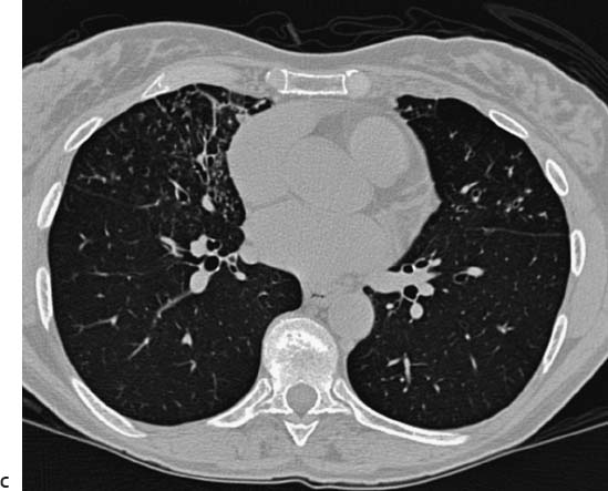
(A) Posteroanterior chest radiograph demonstrates mild interstitial opacity in the medial lower lung fields (arrows). (B) Lateral chest radiograph localizes bronchiectasis and interstitial opacity to the right middle lobe and lingula (arrow). (C) Computed tomography (CT) of the chest (lung windows) demonstrates centrilobular nodules, tree-in-bud opacity, bronchial wall thickening, and bronchiectasis in the right middle lobe (arrow) and lingula.
 Differential Diagnosis
• Nontuberculous mycobacteria (NTMB) infection:
Stay updated, free articles. Join our Telegram channel

Full access? Get Clinical Tree


