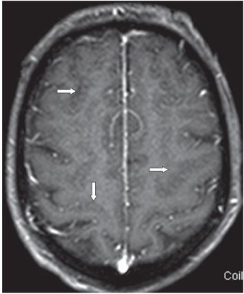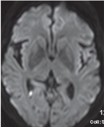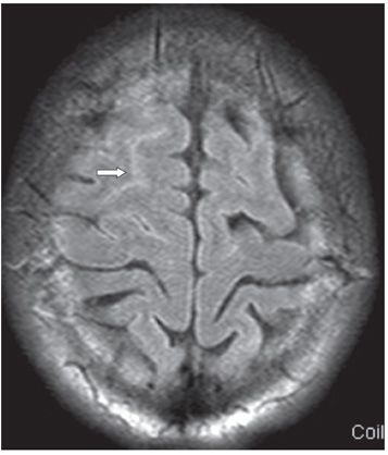


FINDINGS Figure 85-1. Axial FLAIR through the vertex. There is diffuse sulcal hyperintensity over bilateral cerebral hemispheres (arrows point to some of the hyperintense sulci). The sulci appear prominent on T2WI (not shown). Figure 85-2. Axial post-contrast T1WI through same levels as Figure 85-1. There is bilateral sulcal effacement with very little or no leptomeningeal enhancement (arrows). Figure 85-3. Axial DWI through the trigone 2 weeks following antimicrobial treatment. There is a tiny right trigonal restricted diffusion (arrow) compatible with ventricular debris and ventriculitis. Figure 85-4. Axial FLAIR at the same time as Figure 85-3. There is residual right frontal superior sulcus hyperintensity (arrow) with resolution of sulcal hyperintensity and effacement elsewhere.
DIFFERENTIAL DIAGNOSIS Meningitis, subarachnoid hemorrhage (SAH),ventriculitis.
DIAGNOSIS Bacterial meningitis (BM).
DISCUSSION
Stay updated, free articles. Join our Telegram channel

Full access? Get Clinical Tree


