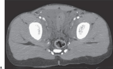
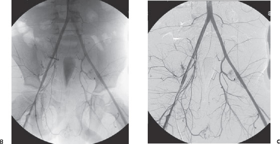
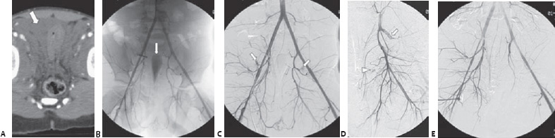
Case 85 A 7-year-old boy struck by a car is brought to the emergency department. (A) Selected pelvic computed tomographic (CT) scan shows hematoma (arrow). (B) Selec ted pelvic angiogram shows teardrop-shaped bladder (arrow) compressed by hematoma. (C) Digital subtraction angiogram shows bilateral hemorrhage from the superior gluteal arteries (arrows). (D) Selected angiogram of the left internal iliac artery after coil embolization of the superior gluteal artery (arrow) shows additional focus of hemorrhage (arrowhead). (E) Pelvic angiogram after Gelfoam embolization of both internal iliac arteries shows no further extravasation.
 Clinical Presentation
Further Work-up
 Imaging Findings
 Differential Diagnosis
 Essential Facts
Stay updated, free articles. Join our Telegram channel

Full access? Get Clinical Tree


