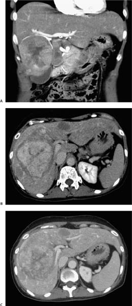
Case 85 A 50-year-old woman presents with abdominal pain and an enlarged liver on physical examination. (A) Coronal, enhanced computed tomography (CT) image in the arterial phase shows a large, heterogeneous mass (arrows) in the right lobe of the liver. The peripheral portions of the mass enhance similarly to liver parenchyma, but the enhancement is variable, with peripheral hyperattenuating and central hypoattenuating regions. (B) Delayed imaging shows the mass to be hyperattenuating compared with normal liver, with delayed enhancement of the central regions (arrowhead). Additional hypoattenuating lesions are visible (arrows) and demonstrate peripherally draped vessels. (C) Portal venous-phase image shows delayed washout of contrast from the bulk of the mass, particularly the central regions (arrowhead). • Hepatic adenoma (telangiectatic):
 Clinical Presentation
 Imaging Findings
 Differential Diagnosis
![]()
Stay updated, free articles. Join our Telegram channel

Full access? Get Clinical Tree


