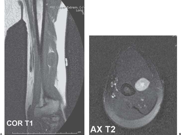
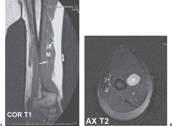
Case 86 A 38-year-old man has a history of a palpable mass in the left arm with paresthesias. (A,B) Coronal T1-weighted magnetic resonance (MR) image of the arm shows a fusiform mass (M) containing homogeneous and isointense signal intensity relative to skeletal muscle. This lesion is located along the course of the radial nerve. A subtle rind of fat is seen at the margins of the mass (arrows). Axial T2-weighted MR image shows a heterogeneous signal increase within the lesion, particularly in the center.
 Clinical Presentation
 Imaging Findings
Stay updated, free articles. Join our Telegram channel

Full access? Get Clinical Tree


